Neurosciences and ophtalmology
From Oct 15th 2003 to Nov 15th 2004, I was a post doc fellow at Steve Shevell's Visual science lab , in the University of Chicago , Chicago, IL - USA.
My research interests inluded
temporal nulling
of chromatic induction, LMS cones sensitivity, receptive field response
according to spatial/temporal modulation, as well as the retinal image
computation
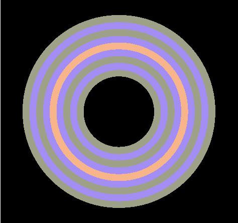
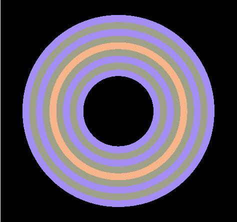
In the above figure, the test circles seems to have different chromaticities (viewed some distance away from the screen ~ 1 meter).
The left one appears orange, whereas the right one appears more pink.
They are physically identical, but due
to the response of a cortical
center / surround spatially antagonistic receptive field, the test
ring's neighborhood induces
this change in color appareance.
Chromatic assimilation and contrast both act to induce this different color perception.
You don't believe me ? Just step closer
to the screen,
and check out by yourself ! (at a viewing distance of a few inches)
During my post-doc
position, I've been
working working on the effects of a
temporal nulling of
such chromatic induction.
A pretty big part of my post-doc researchs focused on the computation of retinal images.
These works are based on
Larry
Thibos's works on the computation of the Point Spread
Function.
The convolution of the point spread
functions by the
wavelength decomposition of the displayed images provides the
wavelength retinal images.
The weighting summation of the wavelength retinal images and the Smith-Pokorny fundamentals gives the L, M & S cones absorption.
The figure below summarizes the different steps of the retinal image computation.
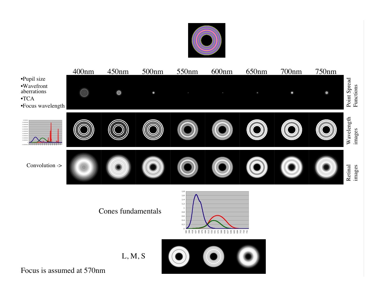
Check out here for further details on the software and its different steps.
I hope I will soon be able to provide some source code and images here !
